Transplant of 'grown' organ
Medical practice
Article on media coverage of an operation to replace a woman's damaged windpipe with one grown in a laboratory from her stem cells.
A 30-year-old Spanish woman has become the “first transplant patient to receive an organ grown to order in a laboratory”, The Independent reported today. It said the woman’s damaged windpipe had been successfully replaced with a “bioengineered organ”. The organ was grown using her own cells on a donor scaffold (a donor trachea stripped of the donor's cells to leave just a cartilage scaffold). In future she will not need to take drugs to suppress her immune system, as is usually required after organ transplant surgery. Extensive media coverage was given to the operation, which The Times newspaper said could “revolutionise” surgery.
This patient will need to be observed to determine the long-term viability of the graft, but the initial results are promising. This technique will now be tried in other patients with similar problems. Further research will be needed to see if the technique can produce other tissues.
Where did the story come from?
Professor Paolo Macchiarini and colleagues from the Hospital Clinic in Barcelona, as well as other research institutions and universities in Spain and the UK carried out this research. The study was funded by the Ministerio de Sanidad y Consumo, Instituto de Salud Carlos III, Fondo de Investigación Sanitaria, Spain; Charles Courtenay-Cowlin Fund, University of Bristol; UK Arthritis Research Campaign; and the James Tudor Foundation. The study was published in the peer-reviewed medical journal The Lancet .
What kind of scientific study was this?
This was a case report that described the transplantation of a tissue-engineered trachea (windpipe) into a patient.
The researchers first developed the technique by experimenting on animals. The process involves the construction of a “tissue-engineered windpipe”, which contains cartilage cells (chondrocytes) grown from the subject’s own stem cells and adhered to a donor windpipe scaffold. By using the subject’s own cells, there is less chance that their immune system will reject the graft.
The researchers had previously managed to generate short, living pieces of trachea in this way, and these had been successfully grafted into animals. Their next step was to attempt to generate a longer piece of living trachea that could be transplanted into a human.
The researchers identified a 30-year old woman who had had various treatments for problems associated with a narrowing of the windpipe leading into her lungs. She had initially suffered from tuberculosis and the condition had eventually led to the removal of part of her windpipe. Scaffolding had then been put in place to hold open the left bronchus (the tube leading from the main windpipe to the left lung).
However, this scaffolding was not well tolerated by the patient’s body, and had to be removed. As a result the bronchus narrowed, her left lung could not function properly and she had severe difficulty breathing. As the only remaining option was to remove the whole left lung, an operation associated with complications and a high death rate, the doctors felt she was a suitable test case. They therefore offered to replace the narrowed part of her bronchus with a tissue-engineered graft.
A 7cm piece of trachea from a deceased female donor was treated to remove all of the donor’s cells, leaving a tubular cartilage scaffold. The researchers then took bone marrow cells and lining (epithelial) cells from the recipient’s bronchus and grew these in the lab. Bone marrow contains stem cells that can develop into any type of cell. In this case, the researchers grew the bone marrow cells in conditions that would lead to them developing into cartilage cells (chondrocytes). The recipient’s chondrocytes and epithelial cells were then ‘seeded’ on to the donor scaffold and allowed to develop in the lab.
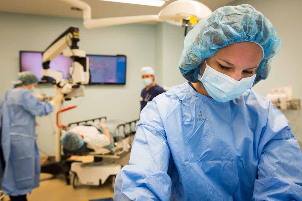
The tracheal graft was then transplanted into the recipient under general anaesthesia. During this procedure, the narrowed part of her bronchus was removed and replaced with a 5cm long piece of the tissue-engineered graft. The patient was monitored after surgery, and tests performed to see if her immune system was producing a response against the donor’s tissue.
What were the results of the study?
The researchers produced a living tissue-engineered graft containing cartilage cells generated from the recipient’s own stem cells and grown on a donor trachea scaffold. The inside of this graft tube was also lined with the recipient’s cells.
This graft successfully replaced a part of the narrowed tube leading from the windpipe into the left lung. The recipient did not experience any complications from the surgery, and was able to leave hospital after 10 days. She was then able to resume normal activities, such as walking up two flights of stairs, walking 500m without stopping, and taking care of her children. Her lung function was normal when tested two months after the surgery. She did not show any immune response against the donor tissue up to two months after surgery and did not need drugs to suppress her immune system.
The graft tissue looked healthy when examined using imaging techniques, and after one month did not look any different to normal tracheal tissue. Cells taken by brushing the surface of the graft after four months also looked normal.
What interpretations did the researchers draw from these results?
The researchers concluded that they could produce a “cellular, tissue-engineered airway” which functions like a normal airway and is free from the risk of rejection. They say their findings suggest that a patient’s own cells, in combination with suitable biological materials, can successfully treat serious medical problems.
What does the NHS Knowledge Service make of this study?
This innovative study shows that it is possible to use a patient’s own cells to reduce the risk of graft rejection. The patient will need to be watched to determine the long-term viability of this graft, but initial results are promising.
This technique will now be tried in other patients with similar problems. Further research will need to determine whether a similar technique can be used to produce other tissues.
Sir Muir Gray adds...
Stem cells have a big contribution to make. This is a very good example of the type of research in which a single case is important.
Article Metadata
Date Published: Mon, 21 Aug 2017


 Subscribe
 Ask the doctor
 Rate this article
 Find products