Eye implant test 'successful'
Medical practice
A miracle eye implant has restored sight to the blind, said the Daily Express. Many newspapers reported this ‘proof of concept’ trial in three patients who were completely blind due to a genetic condition.
A miracle eye implant has restored sight to the blind, said the Daily Express. Many newspapers reported this ‘proof of concept’ trial in three patients who were completely blind due to a genetic condition. Each patient had a microchip implanted into one of their eyes, which was designed to convert light patterns into electrical impulses that could be fed into the optic nerve.
All three patients were better able to perceive light and locate light objects on a dark table. Furthermore, one patient could recognise objects such as a cup and a spoon on a table and could determine letters.
As the Daily Express implies, this is exciting research. Although complete vision restoration remains a long way off, a crude improvement in vision from complete blindness is a promising result. As this was a small pilot study, further work is needed to assess how well the device works in a larger group of patients and to refine the surgical technique and the device itself.
Where did the story come from?
The study was carried out by researchers from The University of Tübingen and other institutes and organisations in Germany and Hungary. The device is produced by Retina Implant AG, Reutlingen, Germany. The trial was funded by the German Federal Ministry of Education and Research. The study was published in the (peer-reviewed) medical journal, Proceedings of the Royal Society B (Biological sciences).
The research was explained well by the newspapers, most of which sensibly mentioned that this is preliminary research in three patients with a particular subtype of blindness and that the vision or light perception gained was modest and not complete.
What kind of research was this?
This clinical pilot study tested whether an experimental device can restore vision in blind adults with a particular form of inherited blindness. The electronic chip that is implanted into the eye is positioned on the damaged retina so that light that enters naturally through the eye’s lens hits the chip. The chip is designed to convert this light into a series of electrical impulses that are picked up by the remaining, undamaged cells in the retina. In theory, these impulses would replace the part of the process of vision that had been damaged by the illness.
The researchers assessed whether the visual function of three blind participants, such as discerning between light and dark and patterns, improved after receiving the implant.
What did the research involve?
The chip had 1,500 individual light-sensitive elements. These were designed to pass on electrical impulses to the nerve cells in the eye. The impulses varied depending on the pattern and intensity of the light that hit the chip.
The study was in two men and a woman aged between 38 and 44 years. All of the patients had hereditary retinal degeneration, but had had good vision prior to losing their site. They had all lost their reading ability at least five years before the study and now only had the ability to perceive light but not to recognise shapes.
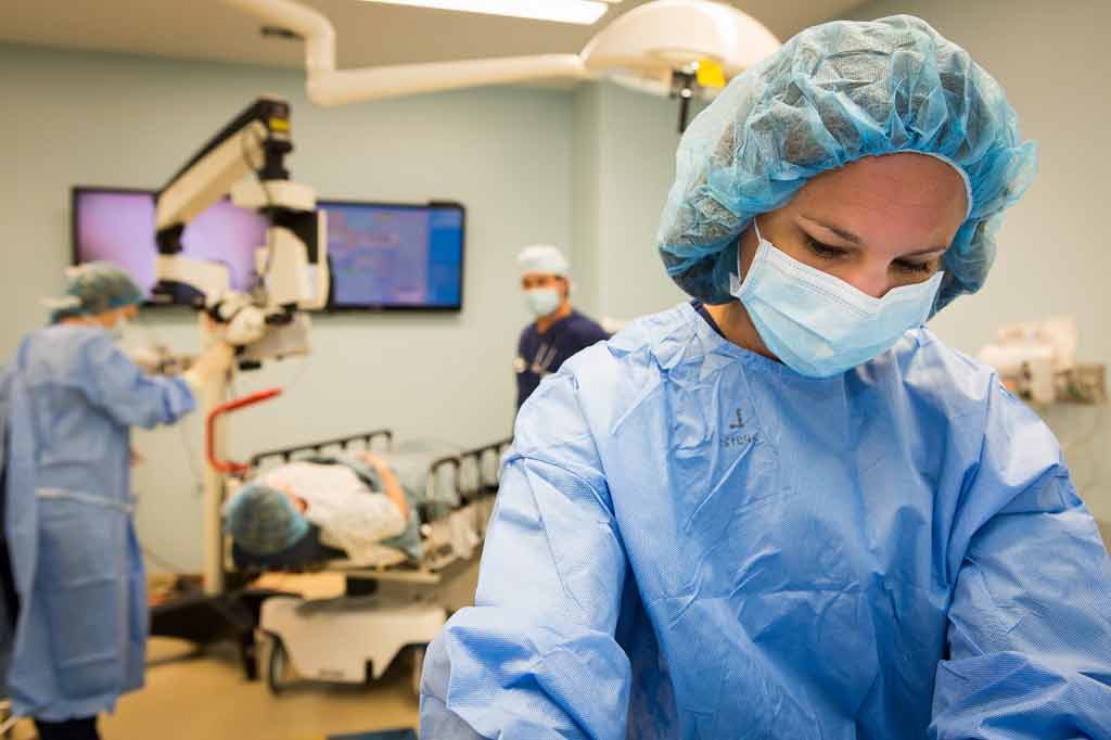
The device was surgically implanted into the eye under the retina. A week later, the patients were given a series of tests of their visual ability to see if they could perceive light, detect movement, and differentiate between different light sources. Tests involved different light stimuli and included being required to identify the direction of some lines (horizontal, vertical of diagonal) and identifying letters and shapes.
What were the basic results?
All three patients were able to perceive light from the chip. Patient two was able to report the direction of the grid lines indicating an improved resolution of light. In the letter recognition task, Patient two was also the only one able to reliably distinguish between different letters, including the letters L, I, T and Z on a screen when the letters were 8.5cm high from a distance of 63cm away. This patient could also differentiate between different shapes and could differentiate seven out of nine contrast differences in a range of grey cards that varied in shade by 15% darkness increments.
In a more natural task, the patients were asked to identify white objects on a black table in front of them. Patient one reliably located a saucer, a square and a cup on the table. Patient three could locate and differentiate a large plate from a saucer. Patient two could locate and correctly describe a spoon, a knife, a cup, a banana and an apple.
The researchers reported that all three of the patients showed distinct learning effects, but that these could not be quantified in this first pilot study.
How did the researchers interpret the results?
The researchers said their study demonstrated that “subretinal micro-electrode arrays can restore visual percepts in patients blind from hereditary retinal degenerations to such an extent that localisation and recognition of objects can provide useful vision, up to reading letters”.
They admit there are still biological and technological obstacles to be overcome and described approaches taken by other groups to develop this sort of device. They said their device had the advantage that all of its parts could be implanted invisibly into the body and could connect with the processing systems of the retina to provide a continuous, stable image.
They say that this study is proof of concept that electronic subretinal devices can potentially improve visual function from a state of complete blindness to one of low vision, thereby allowing localisation and recognition of objects up to reading capability. They say that further development is needed to improve the contrast and spatial resolution that users experience.
Conclusion
This is a proof-of-concept study designed to investigate whether a device of this type could be used to restore any visual function in patients with heredity blindness caused by degeneration of the retina. The research has shown promising results and this was particularly the case in one of the three patients.
The researchers highlight that the patient who had the most successful response was the only one to have had the chip placed under a part of the eye called the macula, the area usually involved in fine central vision. Following this study, further research is needed to optimise the implantation surgery procedure for this device.
Larger studies are now needed to assess how effective this device is and how it can be further improved. The BBC reported that the team are now testing a more compact upgrade to the device, which can be placed entirely under the skin and powered through a socket implanted behind the ear.


 Subscribe
 Ask the doctor
 Rate this article
 Find products


